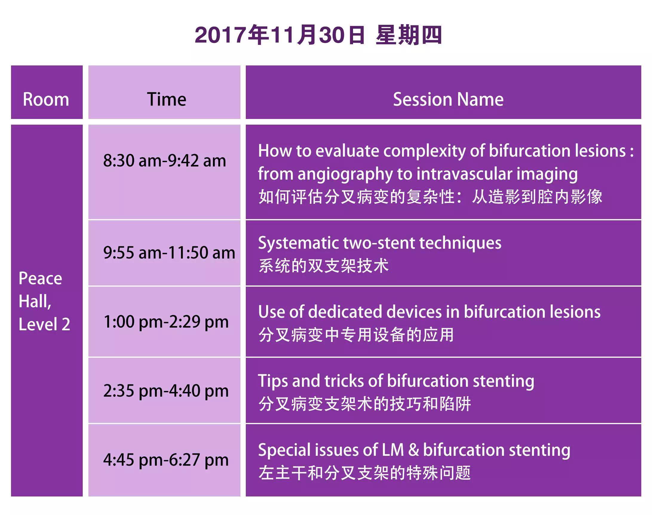
CBS2017 CALENDAR
Event Highlights
IVUS Guidance
Intravascular imaging in coronary artery disease
Mintz GS, Guagliumi G.
Keywords: Intravascular imaging
Although it is the method used by most interventional cardiologists to assess the severity of coronary artery disease and guide treatment, coronary angiography has many known limitations, particularly the fact that it is a lumenogram depicting foreshortened, shadowgraph, planar projections of the contrast-filled lumen rather than imaging the diseased vessel itself. Intravascular imaging-intravascular ultrasound and more recently optical coherence tomography-provide a tomographical or cross-sectional image of the coronary arteries. These techniques are clinically useful to answer questions such as whether the stenosis is clinically relevant; the identification of the culprit lesion; or whether the plaque (or patient) is at high risk of future adverse events. They can also be used to optimise stent implantation to minimise stent-related adverse events, provide answers to the likelihood of distal embolisation or peri-procedural myocardial infarction during stent implantation, and provide reasons for stent thrombosis or restenosis. This review considers the usefulness of intravascular imaging in day-to-day practice.

LEFT MAIN & CORONARY BIFRUCATION SUMMIT
Nanjing Heart Center丨Nanjing First Hospital丨Nanjing Medical University
CHANGLE Rd 68 210006 NANGJING, CHINA
Copyright ⓒ CBSMD Nanjing China. All rights reserved.
FOR REGISTRATION PLEASE CONTACT MS. Ling LIN
+86 25 5227 1398 丨 +86 139 518 845 96 丨 cbs@cbsmd.orgCHANGLE Rd 68 210006 NANGJING, CHINA
Copyright ⓒ CBSMD Nanjing China. All rights reserved.