CBS 2019


CBSMD教育中心




HOME


CBS 2019
At CBS
General Information
Association and Affiliations
Organized by
Nanjing Heart Center
Asian Bifurcation Club (ABC)
Nanjing First Hospital, Nanjing Medical University
Co-organized by
Chinese Society of Cardiology (CSC)
Capacity Building and Continuing Education Center,National Health and Family Planning Commission (CBCEC)
American Society for Cardiovascular Angiography and Interventions (SCAI)
Asian Heart Society (AHS)
Contact Us
Registration Administration of Domestic Representatives
Hongjuan Peng : +86-13913895477
Haimei Xu +86-13914736846
 cbsnj@cbsmd.cn
Registration Administration of Overseas Representatives
Ling Lin : +86 (25) 52271398
+86-13951884596
cbs@cbsmd.cn
Management of Challenging Cases
Yingying Zhao :+86-13815408517
sub@cbsmd.cn
Address
 CBS2019 Congress Secretariat, Nanjing First Hospital, Building No.9 68# Changle Road, Nanjing, China
 210006


FACULTY
FACULTY
General Chairman
Congress Chairman


Education Center
Education Center
• Online Education (For more customized user interface with self-defined collection of research papers and education resources.)
• CBS Education Resource


• CBSMD Scientific Library





SCIENTIFIC LIBRARY
SCIENTIFIC LIBRARY
Research Paper
> Acute Coronary Syndrom
> ASCVD Prevention
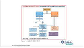
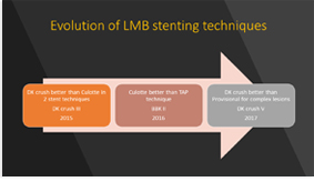
> Bifurcation Stenting
> Cardio-Oncology
> Congestive Heart Failure
> DAPT Duration
> Drug Coated Balloon
> Fractional Flow Reserve
> IVUS Guidance
> Optical Coherence Tomography
> Pulmonary Hypertension
> Rotational Atherectomy
> Shear Stress
> Stenting Left Main
> Transcatheter Aortic Valve Replacement
> Percutaneous LAA Occlusion
> Mitral/Tricuspid Valvular Disease
> Other Relevant Articles
Recommended Paper
Management of Acute Myocardial Infarction During the COVID-19 Pandemic
COVID-19 and Thrombotic or Thromboembolic Disease: Implications for Prevention, Antithrombotic Therapy, and Follow-up
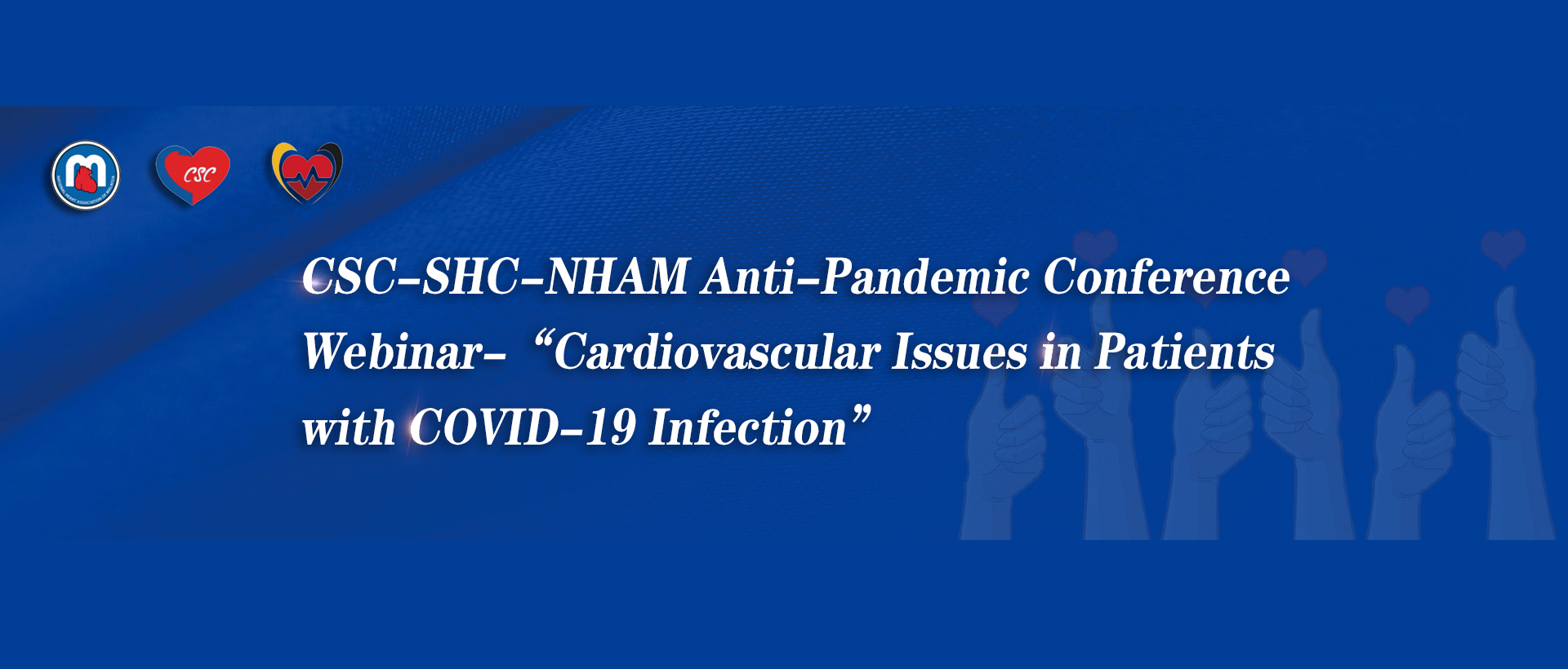
CSC Expert Consensus on Principles of Clinical Management of Patients with Severe Emergent Cardiovascular Diseases during the COVID-19 Epidemic
ACC Clinical Bulletin Focuses on Cardiac Implications of Coronavirus (COVID-19)
光学相关断层扫描
Original Research2018 Feb 15;253:45-49.
JOURNAL:Int J Cardiol. Article Link
La Manna A, Miccichè E, Capodanno D et al. Keywords: Bioresorbable vascular scaffold; Chronic total occlusion; Optical coherence tomography; Qualitative coronary analysis
BACKGROUND - Bioresorbable vascular scaffolds (BVS) have been heralded with potential benefits that are especially desired in long lesions, including chronic total occlusions (CTOs). Procedural feasibility and mid-term outcomes of BVS in CTOs have been reported. However, there is still a paucity of data regarding the vascular and healing response to BVS in CTOs evaluated by optical coherence tomography (OCT).
METHODS - This study included prospectively 21 patients who had a CTO lesion treated with a BVS. Angiography and OCT scan were recorded at either post-implantation and 1-year follow-up. Quantitative coronary angiography and OCT analyses were performed by an independent core laboratory.
RESULTS - The angiographic analysis showed a significant increase in the percentage of in-segment diameter stenosis at 1 year (11.89±9.5% vs. 21.84±11.7%; p=0.002). The OCT analysis showed a trend (p=0.07) towards increased mean scaffold area and significant reductions in mean lumen diameter (3.1±0.36mm vs. 2.85±0.47mm; p=0.0046), mean lumen area (7.8±1.73mm2 vs. 6.76±2mm2; p=0.0082) and minimal lumen area (5.26±1.86mm2 vs. 3.56±1.52mm2; p<0.0001). Malapposition area and volume decreased from 0.26±0.17mm2 to 0.08±0.1mm2 (p=0.0003) and from 14.17±12.92mm3 to 3.99±4.46mm3 (p=0.0014), respectively. The rate of uncovered or malapposed struts, measured at the frame level, was 5.29±6.48% at 1 year.
CONCLUSIONS - In a small series of CTO patients treated with BVS implantation, OCT outcomes at 1year displayed an overall favorable vascular response and healing profile.
Copyright © 2017 Elsevier Ireland Ltd. All rights reserved.






 中 文
中 文




 CBS 2019
 CBS 2019 FACULTY
 Education Resource
 CBSMD Scientific Library


