CBS 2019


CBSMD教育中心




HOME


CBS 2019
At CBS




General Information
Association and Affiliations
Organized by
Nanjing Heart Center
Asian Bifurcation Club (ABC)
Nanjing First Hospital, Nanjing Medical University
Co-organized by
Chinese Society of Cardiology (CSC)
Capacity Building and Continuing Education Center,National Health and Family Planning Commission (CBCEC)
American Society for Cardiovascular Angiography and Interventions (SCAI)
Asian Heart Society (AHS)
Contact Us
Registration Administration of Domestic Representatives
Hongjuan Peng : +86-13913895477
Haimei Xu +86-13914736846
 cbsnj@cbsmd.cn
Registration Administration of Overseas Representatives
Ling Lin : +86 (25) 52271398
+86-13951884596
cbs@cbsmd.cn
Management of Challenging Cases
Yingying Zhao :+86-13815408517
sub@cbsmd.cn
Address
 CBS2019 Congress Secretariat, Nanjing First Hospital, Building No.9 68# Changle Road, Nanjing, China
 210006


FACULTY
FACULTY
General Chairman
Congress Chairman


Education Center
Education Center
• Online Education (For more customized user interface with self-defined collection of research papers and education resources.)
• CBS Education Resource



• CBSMD Scientific Library





SCIENTIFIC LIBRARY
SCIENTIFIC LIBRARY
Research Paper
> Acute Coronary Syndrom
> ASCVD Prevention
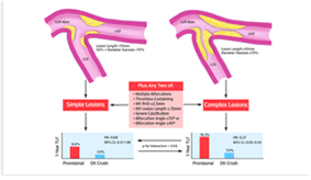
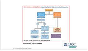
> Bifurcation Stenting
> Cardio-Oncology
> Congestive Heart Failure
> DAPT Duration
> Drug Coated Balloon
> Fractional Flow Reserve
> IVUS Guidance
> Optical Coherence Tomography
> Pulmonary Hypertension
> Rotational Atherectomy
> Shear Stress
> Stenting Left Main
> Transcatheter Aortic Valve Replacement
> Percutaneous LAA Occlusion
> Mitral/Tricuspid Valvular Disease
> Other Relevant Articles
Recommended Paper
Management of Acute Myocardial Infarction During the COVID-19 Pandemic
COVID-19 and Thrombotic or Thromboembolic Disease: Implications for Prevention, Antithrombotic Therapy, and Follow-up
CSC Expert Consensus on Principles of Clinical Management of Patients with Severe Emergent Cardiovascular Diseases during the COVID-19 Epidemic
ACC Clinical Bulletin Focuses on Cardiac Implications of Coronavirus (COVID-19)
Scientific Library
Clinical Trial2019 Nov 2;394(10209):1619-1628.
JOURNAL:Lancet. Article Link
Lanz J, Kim WK, SCOPE I investigators. Keywords: self-expanding vs balloon-expandable bioprosthesis; symptomatic severe aortic stenosis
BACKGROUND - Transcatheter aortic valve replacement (TAVR) is the preferred treatment option for older patients with symptomatic severe aortic stenosis. Differences in the properties of available TAVR systems can affect clinical outcomes. Among patients undergoing TAVR, we compared the self-expanding ACURATE neo TAVR system with the balloon-expandable SAPIEN 3 TAVR system with regard to early safety and efficacy.
METHODS - In this randomised non-inferiority trial, patients (aged ≥75 years) undergoing transfemoral TAVR for treatment of symptomatic severe aortic stenosis, and who were deemed to be at increased surgical risk, were recruited at 20 tertiary heart valve centres in Germany, the Netherlands, Switzerland, and the UK. Participants were randomly assigned (1:1) to receive treatment with the ACURATE neo or the SAPIEN 3 with a computer-based randomly permuted block scheme, stratified by study centre and Society of Thoracic Surgeons Predicted Risk of Mortality (STS-PROM) category. The primary composite safety and efficacy endpoint comprised all-cause death, any stroke, life-threatening or disabling bleeding, major vascular complications, coronary artery obstruction requiring intervention, acute kidney injury (stage 2 or 3), rehospitalisation for valve-related symptoms or congestive heart failure, valve-related dysfunction requiring repeat procedure, moderate or severe prosthetic valve regurgitation, or prosthetic valve stenosis within 30 days of the procedure. Endpoint assessors were masked to treatment allocation. Non-inferiority of ACURATE neo compared with SAPIEN 3 was assessed in the intention-to-treat population on the basis of a risk-difference margin of 7·7% for the primary composite endpoint, with a one-sided α of 0·05. This trial is registered with ClinicalTrials.gov (number NCT03011346) and is ongoing but not recruiting.
FINDINGS - Between Feb 8, 2017, and Feb 2, 2019, up to 5132 patients were screened and 739 (mean age 82·8 years [SD 4·1]; median STS-PROM score 3·5% [IQR 2·6-5·0]) were enrolled. 30-day follow-up was available for 367 (99%) of 372 patients allocated to the ACURATE neo group, and 364 (99%) of 367 allocated to the SAPIEN 3 group. Within 30 days, the primary endpoint occurred in 87 (24%) patients in the ACURATE neo and in 60 (16%) in the SAPIEN 3 group; thus, non-inferiority of the ACURATE neo was not met (absolute risk difference 7·1% [upper 95% confidence limit 12·0%], p=0·42). Secondary analysis of the primary endpoint suggested superiority of the SAPIEN 3 device over the ACURATE neo device (95% CI for risk difference -1·3 to -12·9, p=0·0156). The ACURATE neo and SAPIEN 3 groups did not differ in incidence of all-cause death (nine patients [2%] vs three [1%]) and stroke (seven [2%] vs 11 [3%]); whereas acute kidney injury (11 [3%] vs three [1%]) and moderate or severe prosthetic aortic regurgitation (34 [9%] vs ten [3%]) were more common in the ACURATE neo group.
INTERPRETATION - TAVR with the self-expanding ACURATE neo did not meet non-inferiority compared to the balloon-expandable SAPIEN 3 device in terms of early safety and clinical efficacy outcomes. An early composite safety and efficacy endpoint was useful in discriminating the performance of different TAVR systems.
FUNDING - Boston Scientific (USA).
Copyright © 2019 Elsevier Ltd. All rights reserved.










 中 文
中 文







 CBS 2019
 CBS 2019 FACULTY
 Education Resource
 CBSMD Scientific Library


