CBS 2019
CBSMD教育中心
English

科学研究

科研文章

荐读文献

How to diagnose heart failure with preserved ejection fraction: the HFA-PEFF diagnostic algorithm: a consensus recommendation from the Heart Failure Association (HFA) of the European Society of Cardiology (ESC) Six Versus 12 Months of Dual Antiplatelet Therapy After Implantation of Biodegradable Polymer Sirolimus-Eluting Stent: Randomized Substudy of the I-LOVE-IT 2 Trial Effect of Intravascular Ultrasound-Guided vs Angiography-Guided Everolimus-Eluting Stent Implantation: The IVUS-XPL Randomized Clinical Trial Ambulatory Inotrope Infusions in Advanced Heart Failure - A Systematic Review and Meta-Analysis Cardio-Oncology: How New Targeted Cancer Therapies and Precision Medicine Can Inform Cardiovascular Discovery Catastrophic catheter-induced coronary artery vasospasm successfully rescued using intravascular ultrasound imaging guidance Initial Invasive or Conservative Strategy for Stable Coronary Disease Positive recommendation for angiotensin receptor/neprilysin inhibitor: First medication approval for heart failure without "reduced ejection fraction" Nitrosative stress drives heart failure with preserved ejection fraction Combined Tricuspid and Mitral Versus Isolated Mitral Valve Repair for Severe MR and TR: An Analysis From the TriValve and TRAMI Registries

Original Research

JOURNAL:ACC Article Link

TAVR: Role of Multimodality Imaging

Pre-reading

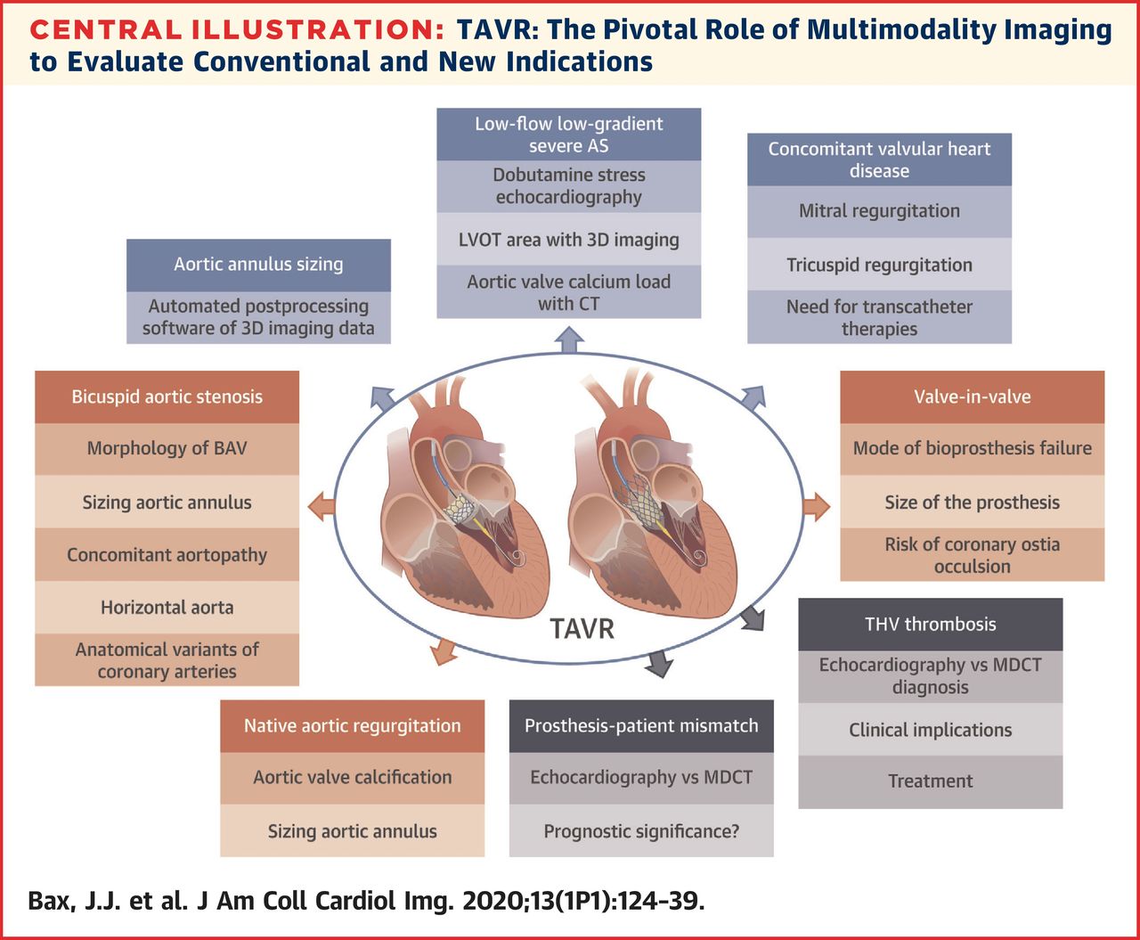
The following are key points to remember from this state-of-the-art review on transcatheter aortic valve replacement (TAVR) and the role of multimodality imaging in common and complex clinical scenarios:

  1. 1. TAVR has rapidly become an established therapy for patients with symptomatic severe aortic stenosis (AS).
  2. 2. Technological advances and the learning curve have resulted in better procedural results in terms of hemodynamic valve performance and intermediate-term clinical outcomes.
  3. 3. The integration of anatomical and functional information provided by multimodality imaging has improved size selection of TAVR prostheses, permitted better patient selection, and provided new insights in the performance of the TAVR prostheses at follow-up.
  4. 4. The use of 3D imaging techniques (multi-detector row computed tomography [MDCT], cardiac magnetic resonance [CMR], and 3D echocardiography) that permit accurate measurement of the left ventricular outflow tract area by direct planimetry has demonstrated the ability to reclassify severe AS patients into moderate AS by 12% in patients with low-flow, low-gradient severe AS.
  5. 5. Furthermore, the field of TAVR continues to develop and expand the technique to younger patients with lower risk on the one hand, and more complex clinical scenarios, on the other hand, such as degenerated aortic bioprostheses, bicuspid aortic valves, or pure native aortic regurgitation.
  6. 6. The use of both echocardiography and MDCT is key in the diagnosis of patients with severe AS who may benefit from TAVR as well as in the procedural planning and evaluation of the results at follow-up.
  7. 7. The number of patients with bicuspid AS treated with TAVR is increasing and the TAVR results with the use of new generation prostheses are promising.
  8. 8. TAVR in degenerated bioprosthesis has been an important recent breakthrough because re-operation in these individuals is associated with very high mortality.
  9. 9. Patients with native aortic regurgitation are also now being treated with TAVR.
  10. 10. These newer indications for TAVR need careful imaging evaluation of the anatomy of the landing zone to ensure successful anchoring of the TAVR prosthesis and to minimize complications. These new horizons for TAVR are making multimodality imaging critically important for this evolving therapy.